Language switcher


Video Microscopy
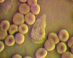
Capillary blood as seen with a video microscope: erythrocytes and granulocytes in action
Thanks to the constant development and improvement of scientific instruments such as video microscopes, it is now possible to study living cells in their real environment, instead of observing dead, dyed cells.
Structures are formed by processes. Today we strive to study and understand living processes in the context of their environment, and to influence it in a positive way.
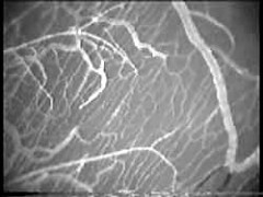
A video image of the terminal circulation (living bone)
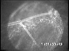
With vital staining, the cells in flowing blood become clearly visible. The photo is obtained from same sample as on the left-hand side – living bone.